Fig. 6
From: Masses of developmental and genetic origin affecting the paediatric craniofacial skeleton

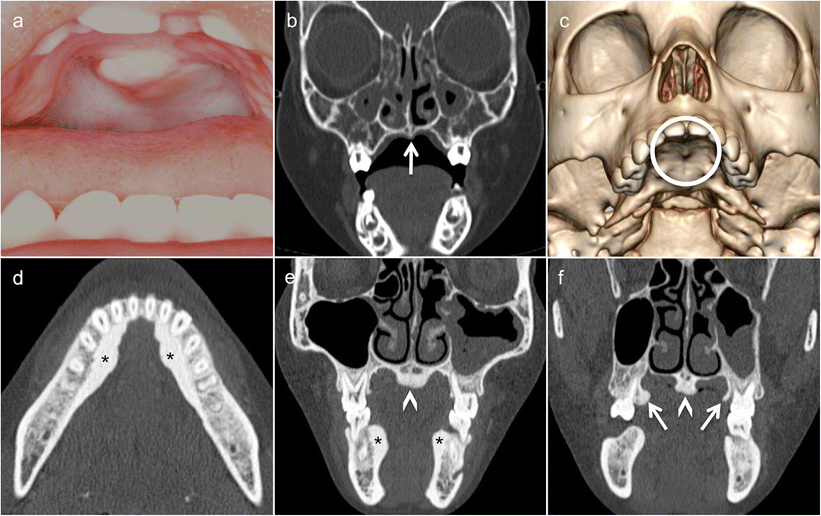
Torus palatinus (TP) in a 3-year-old girl who underwent imaging because of an indurated palpable midline mass of the hard palate increasing in size (a) after a fall occurring from a swing hanging on a tree two weeks earlier. Coronal low-dose CT image (bone window) (b) and three-dimensional CT VR reconstruction show a small midline spur/exostosis (arrow and circle). Characteristic aspect of a torus mandibularis (TM) (asterisks in d and e), of a TP (arrowheads in e and f) and of a torus maxillaris (TMax) (arrows in f) as seen in a young adult. It is worthwhile mentioning that TM and TMax are extremely rarely diagnosed in children