Fig. 11
From: MRI of the axial skeleton in spondyloarthritis: the many faces of new bone formation
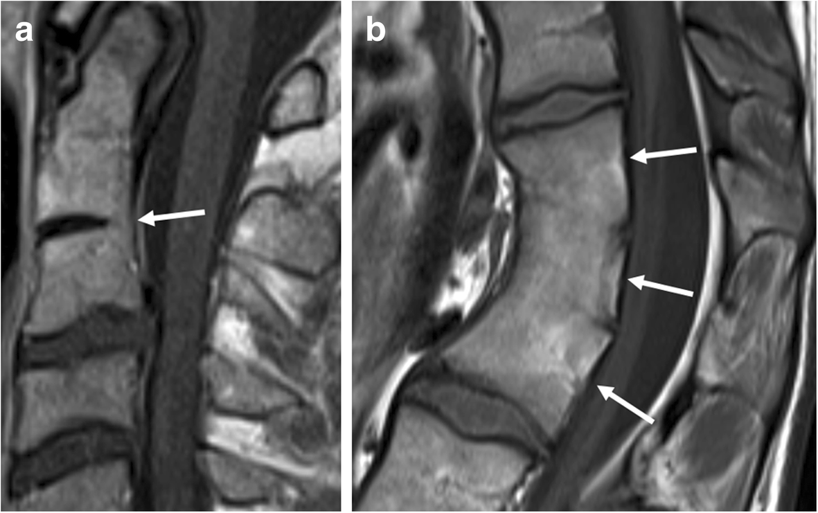
Pitfalls in imaging and diagnosis of new bone formation: congenital block vertebrae. Sagittal T1-weighted MR images show (a) partial congenital block vertebra of C2-C3 (arrow) and (b) complete congenital block vertebra consisting out of three vertebrae (arrows). Note the narrowed antero-posterior diameter in b, a typical sign of complete congenital fusion. Also, note that the height in b is less than the expected sum of three vertebrae and two intervertebral discs