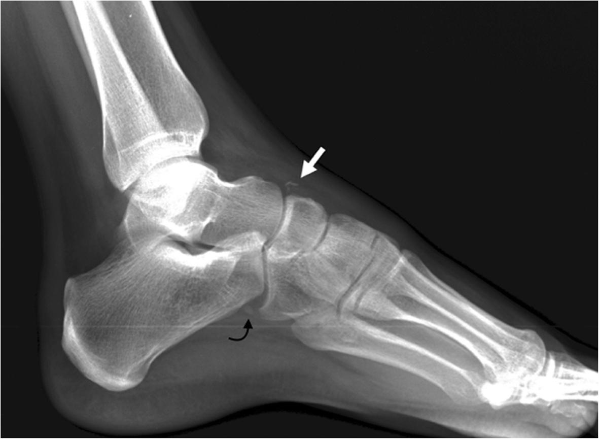
Fig. 7

Differential diagnosis of os supranaviculare. The differential diagnosis has to be established with an avulsion fracture of the talo navicular joint capsule, as seen in this lateral radiograph (white arrow). Note the mild swelling of the soft tissues associated and the more linear configuration of the fragment, compared to the more triangular shape of the ossicle. Incidental note os peroneum (curved black arrow)